
Advances in Animal Orthopedics: Fixing Wings and Limbs
Published on December 26, 2025 by Admin
Introduction
Veterinary orthopedic surgery has advanced significantly. This progress allows us to treat complex bone and joint issues in animals. These advancements are crucial for both pets and wildlife. This article explores these exciting developments.
Understanding Orthopedic Conditions in Animals
Orthopedic surgery addresses issues with bones, joints, ligaments, and muscles. Animals can experience pain from various conditions. These include joint problems, torn ligaments, and broken bones. Congenital issues can also be corrected. For instance, the cranial cruciate ligament (CCL), often called the anterior cruciate ligament (ACL) in humans, is a common focus. These problems can cause significant discomfort and mobility loss.
Recognizing the Signs
Pay close attention to how your pet moves. Unusual changes in gait can indicate an orthopedic problem. Symptoms often include difficulty standing up. They might favor one leg intermittently. Limping is another common sign. Swelling in a limb or stiffness can also occur. A decreased activity level is also a key indicator.
If you notice any of these signs, seek veterinary care promptly. Early diagnosis leads to better outcomes. A thorough examination is essential. Veterinarians can then recommend the best course of treatment.
Surgical Interventions for Fractures
Fractured bones are a primary target for orthopedic surgery. Treatment methods vary based on the animal, fracture type, and location.
Repairing Broken Bones
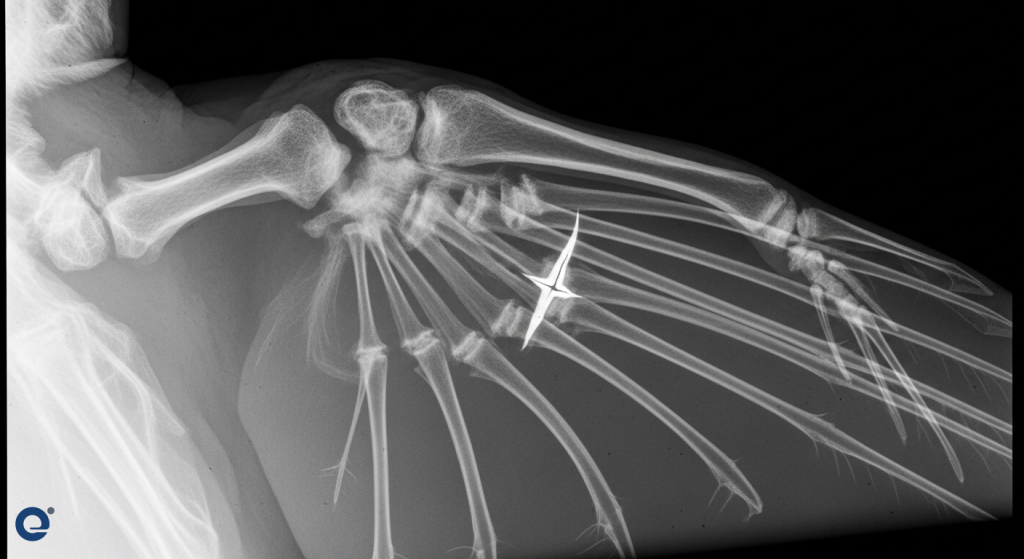
Veterinary surgeons can repair fractured bones using several techniques. These include pinning and external fixator placement. Pinning involves inserting a metal pin into the bone. This stabilizes the fracture fragments. External fixators use pins that go through the skin and bone. These pins are connected to an external frame. This method provides excellent stability.
For example, a red-tailed hawk suffered a mid-shaft oblique fracture of its humerus. The veterinary team opted for surgery using a pin and wires. This approach is preferred for birds due to their lightweight bones. Plates and screws can be too heavy and may require removal later. The pin is easily removed without anesthesia after healing. The wires offer stability and are light enough not to impede flight.
In another case, an ostrich named Twig at the Hattiesburg Zoo sustained a broken proximal humerus in her wing. LSU Vet Med surgeons repaired this clean break with minimal displacement. They used a permanent plate and screws for long-term stability. This procedure allowed Twig to recover and regain function in her wing.
Advanced Fracture Repair Techniques
New technologies are continually improving fracture repair. For complex pelvic fractures, such as sacroiliac luxation/fracture in small dogs, specialized tools are used. The SILIS-MILAD system, for instance, allows for minimally invasive repair. This technique uses a scope to guide screw placement. It significantly reduces surgery time and trauma. A dog named Biscuit, injured in a traffic accident, had his pelvic bones repositioned and secured with lag screws using this method. The surgery took only 17 minutes, a stark contrast to the 45-90 minutes of an open approach.
Addressing Ligament and Joint Injuries
Ligament injuries, particularly in the knee, are common. The cranial cruciate ligament (CCL) is a critical stabilizer in a dog’s knee. Rupture of the CCL is a leading cause of hind limb lameness and pain.
Canine Cruciate Ligament Disease
Unlike in humans, CCLD in dogs is often due to degeneration. Factors like aging, obesity, poor conditioning, and conformation contribute. This means the ligament gradually weakens over time. Consequently, partial tears can progress to complete ruptures. This degeneration often affects both knees. Therefore, dogs with a CCL injury in one leg have a high risk of developing it in the other.
Symptoms include difficulty rising, altered sitting posture, and decreased activity. A popping sound might indicate a meniscal tear. Swelling and muscle atrophy can also be present. Surgical repair aims to restore joint stability and reduce pain. Various surgical techniques exist, such as tibial plateau leveling osteotomy (TPLO) and extracapsular repair. These procedures help manage pain and slow arthritis progression. Regular exercise and weight management are vital for prevention and recovery.
While CCLD is common in dogs, cats are rarely affected. Certain breeds like Rottweilers and Labrador Retrievers have a higher incidence. Female and neutered dogs also appear to be at greater risk.
Specialized Procedures for Exotic and Wild Animals
Orthopedic surgery is not limited to domestic pets. Exotic animals and wildlife also benefit greatly from these advancements.
Wing and Limb Amputations
In severe cases, amputation of a limb or wing may be necessary. This can be a life-saving procedure. It allows animals to adapt and survive, especially in wildlife rehabilitation settings. Amputation can provide a better quality of life than prolonged suffering from an irreparable injury.
Other Specialized Surgeries
Beyond fractures and ligament repairs, veterinary orthopedic surgeons perform other specialized procedures. These can include femoral head osteotomies, beak repair, and chelonian (turtle/tortoise) shell repair. These procedures require a deep understanding of the unique anatomy and physiology of different species. Collaboration with board-certified veterinary surgeons is common for complex cases.
The Role of Technology and Innovation
Technological advancements are revolutionizing animal orthopedic surgery. Minimally invasive techniques reduce patient trauma and recovery times.
Imaging and Diagnostics
Advanced imaging, including CT scans and high-resolution X-rays, is crucial. These tools provide detailed views of bone and joint structures. They enable precise diagnosis and surgical planning. For instance, preoperative CT scans were vital in assessing the extent of pelvic fractures in Biscuit. This allowed for accurate pre-operative planning.
Surgical Tools and Implants
New surgical instruments and implants enhance surgical precision and outcomes. The SILIS-MILAD system exemplifies this innovation for pelvic surgeries. Implants are increasingly designed for specific species and anatomical locations. Biocompatible materials are used to ensure good integration with the animal’s body. The goal is always to restore function and minimize long-term complications.
The Surgical Process and Recovery
A successful orthopedic surgery involves meticulous pre-operative care, skilled execution, and diligent post-operative management.
Pre-Operative Care
Before surgery, animals undergo thorough examinations. This includes bloodwork and imaging. Pain management is initiated. For wildlife, stabilization and supportive care are provided to ensure they are strong enough for anesthesia. For example, the red-tailed hawk received pain medication, anti-inflammatories, assist feeding, and fluids to prepare for surgery.
The Surgical Procedure
During surgery, anesthesia is carefully managed. The surgical site is prepared aseptically. The chosen surgical technique is then performed. Surgeons use fluoroscopy (real-time X-rays) to guide instrument placement during some procedures. This ensures accuracy, especially in complex repairs like the SILIS-MILAD surgery.
Post-Operative Care and Rehabilitation
Recovery is a critical phase. Post-operative care includes pain management, wound care, and restricted activity. Rehabilitation often involves physical therapy. This helps restore strength, flexibility, and function. For wildlife, the ultimate goal is often a return to their natural environment. This requires successful healing and regained mobility. Twig, the ostrich, is monitored daily for signs of discomfort. Her recovery involves regular X-rays to assess healing.
Challenges and Future Directions
Despite advancements, challenges remain in animal orthopedic surgery. Cost, availability of specialized equipment, and species-specific anatomical differences present hurdles.
Species-Specific Needs
Caring for exotic and wild animals presents unique challenges. Their delicate bone structure, physiological differences, and stress responses require specialized knowledge. For example, the choice between pins and wires versus plates and screws for birds highlights these species-specific considerations.
The Future of Orthopedic Veterinary Medicine
The field continues to evolve. Research into regenerative medicine, advanced implant materials, and robotic-assisted surgery holds promise. Furthermore, telemedicine can expand access to specialized orthopedic consultations for remote clinics. The focus remains on improving animal welfare, restoring function, and enhancing the quality of life for all animals.
Frequently Asked Questions
What are the main goals of orthopedic surgery in animals?
The primary goals are to repair fractured bones, correct joint problems, stabilize torn ligaments, and address congenital deformities. Ultimately, the aim is to alleviate pain, restore function, and improve the animal’s quality of life.
How do veterinarians diagnose orthopedic conditions?
Diagnosis typically involves a thorough physical examination, observing the animal’s gait and range of motion. Imaging techniques like X-rays, CT scans, and MRIs are essential for visualizing bone and soft tissue structures. Blood tests may also be performed to assess overall health and rule out other conditions.
What is the recovery process like after orthopedic surgery?
Recovery varies greatly depending on the procedure and the animal. It generally involves pain management, restricted activity, and often physical rehabilitation. Strict adherence to post-operative instructions is crucial for successful healing. For wildlife, rehabilitation aims to return them to their natural habitat.
Can all orthopedic injuries in animals be surgically repaired?
While many injuries can be effectively treated, some may be too severe or complex for surgical repair. In such cases, palliative care or amputation might be considered to ensure the animal’s comfort and well-being. The decision is always based on the individual animal’s condition and prognosis.
Conclusion
Advances in orthopedic surgery are transforming animal care. From mending the broken wings of wild birds to restoring mobility in companion animals, these techniques offer hope and healing. The dedication of veterinary surgeons and the integration of new technologies ensure a brighter future for animal orthopedics. This ongoing progress allows more animals to lead pain-free, functional lives.